Why is physics beautiful?
The physics done at CERN aims at understanding how particles and forces act in the subatomic world and are organized to form everything we observe around us. By using what we learn on the path towards larger and larger collision energies, we also model the first instants after the Big Bang, thus answering fundamental questions about the coming into existence of the Universe.
I think that nobody can remain unmoved by the inner beauty of the world and thus of the science that allows us to perceive it in all its depth.
2014 marks the 50th anniversary since the discovery of quarks, 30 years from the discovery of the carriers of the electroweak interaction here at CERN and the second anniversary since the discovery of the Higgs field, which so magnificently complete and crowns the Standard Model of particle physics. In that sense, the beginning of 2014 is the proper time to first describe these “beautiful” physics results, together with their consequences in our description of the events that took place in the first millionth of a second of the Universe life.
In addition, the development of CERN basic research in particle physics is closely linked to useful applications that have greatly changed our lives. This is no coincidence: indeed, the beauty of particle physics has always been going hand in hand with useful applications.
It is clear to anybody who visits a hospital that physics applications are everywhere. We use physics when we measure the blood pressure, when we perform an ultrasound scan to determine the sex of an unborn child, when we take radiography or a CT scan.

Image is taken from a recent video showcasing particle therapy, an advanced technique of cancer radiation therapy using protons or other ions, watch the video:http://cds.cern.ch/record/1611721
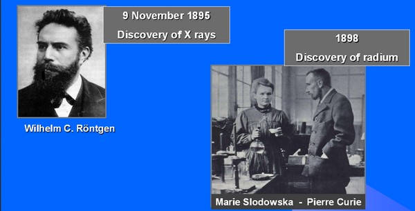
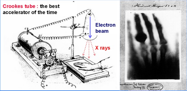
How did this useful application of physics come about? If you look at 120 years of history by following the tracks of particle accelerators, you will find that most of these applications were not thought of when people discovered a new phenomenon or invented a new type of instrument. They themselves were surprised by the applications. The first example is the discovery of X-rays in 1895 by Roentgen, which started modern fundamental physics and was used within months, all over the world, for the radiography of bones and of wounded soldiers. The first use in cancer therapy of X-rays and radium – discovered in 1898 by Marie and Pierre Curie - was also almost immediate. It all started in January 1895 when Emile Grubbe, a student in the University of Chicago, used X-rays to treat a breast cancer; though the patient died a few weeks later it opened the way to the possible uses of X-rays for medical treatments.
The beginning of fundamental and medical physics
First medical use of an accelerator
These events point to the picture of three intertwined yarns. The first is fundamental physics (black), the second is therapy (red like blood) and the third is diagnostics (blue), the art of diagnosing medical problems with the highest precision possible and on time to cure the disease and avoid further complications. These three yarns are strongly intertwined and along them we can put all the major events that happened along the years one traces the history of ideas, people, inventions and discoveries from one yarn to the other.
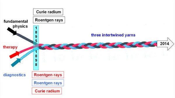
Three intertwined yars showing 120 years of beautiful and fundamental physics.
Perhaps one of the most important steps that cut across the three yarns is the building of the first cyclotron. In 1932, Lawrence commissioned in Berkeley its first cyclotron, which he invented a few years earlier in Berkeley. He and his collaborators immediately discovered that the radioisotopes, which were produced by this new type of accelerator in abundance, were useful for doing both diagnostics, i.e. following the substances in the body of the patient, and for therapy. For instance, his brother John Lawrence, who was a medical doctor, injected radioactive phosphorus 32 in the body in order to treat leukaemia. In 1939, the 60-inch cyclotron was financed exclusively for medical purposes and later was used not only for research in nuclear physics but also to treat patients with neutron beams; another great example of how developments along the three yarns are correlated.
The same applies for the invention of the synchrotron that was made possible thanks to the “phase stability principle” proposed independently in the States and in Soviet Union by E. McMilan and V.J.Veksler. LHC is in made of two interlinked synchrotrons and the centre - which in Pavia is treating radioresistant tumours with beam of carbon ions - was designed by the TERA Foundation on the basis of the Proton Ion Medical Machine Study carried out at CERN in the years 1996-2000 under the leadership of Phil Bryant.
Nowadays we see the interdependence of the three yarns as 10 MeV electron linear accelerators – similar to the one that was injecting electron and positrons in LEP - dominate in “conventional radiotherapy: in the world their number peaks at 20 000 and their total length equals the length of LHC. Moreover for fifteen years TERA has been developing novel hadron linacs for proton therapy, the tumour treatment that spares healthy tissues better than X-rays.

The invention and development of synchrotron have pushed modern high energy physics and contributed to the development of medical applications. CNAO accelerator (photo courtesy: CNAO)
Thus synchrotrons and linacs have pushed modern particle physics over the last 50 years to the frontier that we are standing today and are used around the world in many thousands therapy medical centres. CERN has also contributed to the early development of tools for better diagnostics. Indeed, Alan Jeavons and David Townsend made the first diagnostic application, using the first PET detector based on the wire chambers invented by George Charpak and developing special software, they took the first mouse image with the participation of radiobiologist Marilena Streit-Bianchi in 1977 at CERN.
Looking back to this long story, I am very happy that Rolf Heuer, CERN’s DG, got the approval of the CERN Council for the creation of a new office dedicated to medical applications. Steve Myers, one of the leading figures of the LHC developments, has been put in charge. I think the establishment of the new office is very promising for the development of useful physics at CERN for the years to come.