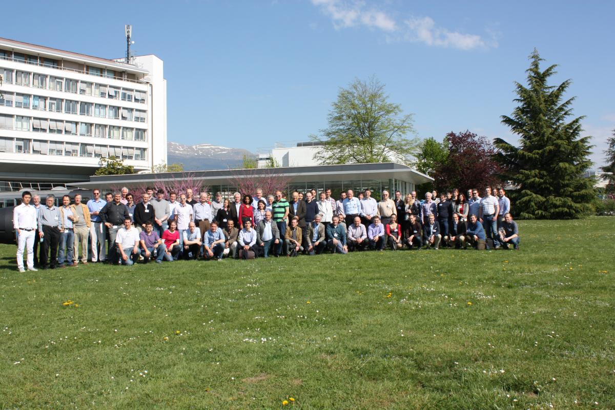
3rd workshop on medical applications of spectroscopic X-‐ray detectors.
The 3rd workshop on medical applications of spectroscopic X-ray detectors took place at CERN from 20-23 April. There were 113 registered participants of which about 50 were from industry. All of the major medical equipment suppliers active in the spectroscopic Computer Tomography (CT) field (GE, Philips, Siemens and Toshiba) sent significant delegations. There was also a sizable participation from some of the most prestigious medical schools (Johns Hopkins, University of Massachusetts, Mayo Clinic, Royal Marsden, TU Munich, etc.).
Dr Sahani, a leading radiologist from Massachusetts General and Harvard Medical School, presented the workshop keynote talk. The state of the art in clinical practice is dual-energy CT. He reported that 2/3 of all CT’s now use this modality as it gives a significant improvement in diagnostic precision without adding dose. In the round table discussion which followed the industry session (which included presentations from the big 4 equipment suppliers) it became clear that each have major internal efforts dedicated to exploiting the new modality, which permits more information to be extracted from the dose to which the patient is subjected. In a session dedicated to new biomarkers there was quite some optimism that functional information may become accessible using CT, in some cases obviating the need for more expensive MRI or PET examinations.
As well as sessions on reconstruction algorithms, prototype imaging systems and imaging performance there were quite a number of papers from companies which have grown out of HEP developments. These companies have often targeted the synchrotron market to begin with but are now moving towards medical applications. The last regular sessions were devoted to high-Z semiconductors and readout ASICs. CdTe and CZT are now reported to be able to deal with the fluxes required by medical CT. The readout ASICs are, of course, a key element in the system and significant progress was reported.
Moreover a number of CERN-related topics were presented during the workshop. Rafa Ballabriga presented a review of the currently available ASICs highlighting some important trends. He showed that the highest count rates can be obtained with small pixel pitches but then the use of charge summing and hit allocation (first deployed in Medipix3) is essential if useful spectroscopic information is to be retained. Erik Frojdh (CERN Ardent student) presented the Timepix3 chip which could become an important tool in understanding the charge deposition process in detector materials, thereby helping in the optimisation of new ASIC readout architectures. Steve Myers welcomed the delegates to CERN on the first day and gave a presentation overviewing CERN medical applications with an emphasis on hadron therapy. Jim Virdee gave a much-appreciated talk in the closing session. The workshop summary talk was given by Anthony Butler, a radiologist from Christchurch (NZ) and a member of the team developing the MARS Medipix3-based scanner.
There was a lot of enthusiasm from the delegates because of the sharp focus of the workshop and the format which permitted deep discussions and interactions between the experts in this very specific field.