Colour X-rays for Medicine: The 2019 specXray workshop

The fifth workshop on Medical Applications of Spectroscopic X-ray Detectors (specXray) took place at CERN from 13 to 17th of May. Since its establishment in 2011, the workshop has brought together specialists from different but related fields focusing on how best to advance effort and understanding in the new imaging modality of spectroscopic X-ray imaging. The 120 participants comprised clinicians, radiologists, biologists, tracer developers, imaging system specialists and detector and ASIC developers.
All of the major medical radiology equipment suppliers were represented along with researchers from many of the major medical schools. Prof Anthony Butler from the University of Canterbury (New Zealand) in the closing summary of the conference noted: “Since the first meeting, scepticism about the benefits of such an approach (and the associated technical challenges) has gradually given way to an acceptance that the approach has great potential, offering new avenues in diagnostic imaging.”
Indeed, since the discovery of the x-rays by Röntgen in 1895, there is a continuing interplay between physics and medicine boosting both imagining and treatment techniques. More specifically, in the case of medical imaging, technological advancements for particle detectors have fuelled new developments in medical imaging.
A number of image metrics, presented by clinicians during the workshop together with practical case studies carried out using prototype clinical systems have shown that spectroscopic photon counting gives better image quality at reduced dose. Moreover, studies have shown that multiple contrast agents can be distinguished leading to potential applications in functional X-ray imaging.
Perhaps it should be noted that last year, scientists in New Zealand applied the spectroscopic imaging technique to perform the first-ever 3-D, colour X-ray on a living human using a Medipix3-based system, promising to revolutionize the field of medical diagnostics. The latest results were presented by Butler and first clinical trials are underway.
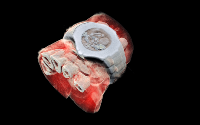
A 3D image of a wrist with a watch showing part of the finger bones in white and soft tissue in red. (Image: MARS Bioimaging Ltd)
The mood of the meeting was rather positive with most participants convinced that spectroscopic X-ray imaging will find its place in clinical practice in the coming years. “This is what happens when you bring people with good ideas together to form a community” noted Stephan Kappler, a member of the Scientific committee. With more than 300,000,000 CT scans being performed annually around the world the potential impact for the spectroscopic X-ray imaging modality is huge.
Workshop webpage: http://specxray.web.cern.ch/specxray/
You can find more information about the Detector Seminar: https://indico.cern.ch/event/819651/